The development of effective high-throughput tools in drugĭiscovery research has increased the demand for complementary high capacity immunoblot methods in which to assess the consequences of drugĬandidates at protein level. Because of its prevalence and the severity of the clinical manifestations, finding a cure for DM1 is a social and medical need. In its most common form, the onset of symptoms occurs during adolescence, and affected people have a significantly shortened lifespan of 48–55 years.

Currently, there is no effective treatment for DM1, and management of symptoms is the only option to preserve the quality of life of people living with DM1. Ītrophy and weakness, which, in advanced stages of the disease, lead to respiratory distress and early death. Studies in animal models have shown that the increase of MBNL in a geneticīackground of DM improves the pathological phenotypes and that the overexpression of MBNL1 in control mice is well tolerated. The depletion in MBNL protein function causes alterations in RNA metabolism that originate defined symptoms of the disease. CUG expansion RNA aberrantly binds and sequesters essential developmental proteins of the Muscleblind-like (MBNL) family, which are key regulators of alternative splicing. DM1 originates from an expansion of the CTG trinucleotide repeat in the 3′-untranslated region (UTR) of the DMPK gene that, upon transcription, formsĬUG hairpins that behave as toxic RNAs. For chemiluminescence signal detection, apparatus need to be disassembled and the membrane need to be taken out and wrapped in a transparent plastic film.Myotonic dystrophy type 1 (DM1) is a degenerative geneticĭisease that is classified as rare because it affects less than 1 in 2000 people (1/3000 to 1/8000 ). Vacuum-assisted dot blot apparatus has been used to facilitate the rinsing and incubating process by using vacuum to extract the solution from underneath the membrane, which is assembled in between several layers of plates to ensure good seal between sample wells, hold waste solution, and deliver suction force. Finally, for chemiluminescence imaging, the piece of membrane need to be wrapped in a transparent plastic film filled with enzyme substrate. After the protein samples are spotted onto the membrane, the membrane is placed in a plastic container and sequentially incubated in blocking buffer, antibody solutions, or rinsing buffer on shaker. After antibody binding, the membrane is incubated with a chemiluminescent substrate and imaged.ĭot blot is conventionally performed on a piece of nitrocellulose membrane or PVDF membrane. It is then incubated with a primary antibody followed by a detection antibody or a primary antibody conjugated to a detection molecule (commonly HRP or alkaline phosphatase). The membrane is incubated in blocking buffer to prevent non-specific binding of antibodies. Samples can be in the form of tissue culture supernatants, blood serum, cell extracts, or other preparations. Methods Ī general dot blot protocol involves spotting 1–2 microliters of a samples onto a nitrocellulose or PVDF membrane and letting it air dry. Performing a dot blot is similar in idea to performing a western blot, with the advantage of faster speed and lower cost.ĭot blots are also performed to screen the binding capabilities of an antibody. However, it offers no information on the size of the target protein.

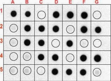
The technique offers significant savings in time, as chromatography or gel electrophoresis, and the complex blotting procedures for the gel are not required. Instead, the sample is applied directly on a membrane in a single spot, and the blotting procedure is performed. It represents a simplification of the western blot method, with the exception that the proteins to be detected are not first separated by electrophoresis. Darker dots indicate more protein.Ī dot blot (or slot blot) is a technique in molecular biology used to detect proteins.


 0 kommentar(er)
